Healthcare facilities all over the world use Woods lamps, also known as fluorescent spectroscopy, ultraviolet light to visually diagnose a wide variety of bacterial and fungal skin infections, skin pigment disorders, and other irregularities. Woods lamps use ultraviolet light to reveal skin abnormalities that can’t be seen with human eyes. For dermatologists, visual diagnoses are imperative, and Woods lamp testing is an essential step in the patient evaluation process. Woods lights and woods lamps both refer to the same item.
Top 9 Medical Diagnostic Applications for a Woods Lamp
A Woods lamp can help diagnose a variety of infections and conditions, including the following:
1. Tinea Capitis
2. Vitiligo
3. Corneal Abrasions
4. Ringworm
5. Porphyria
6. Pigment Disorders
7. Bacterial Infections
8. Acne Vulgaris
9. Other Miscellaneous Uses
By using black light to illuminate areas of the skin surface, medical professionals can easily see the presence of bacteria and fungi, which cause affected skin areas to change color under the light. In fact, the use of ultraviolet light can reveal a great deal of information about both the physical and the chemical properties of a patient’s skin and hair.
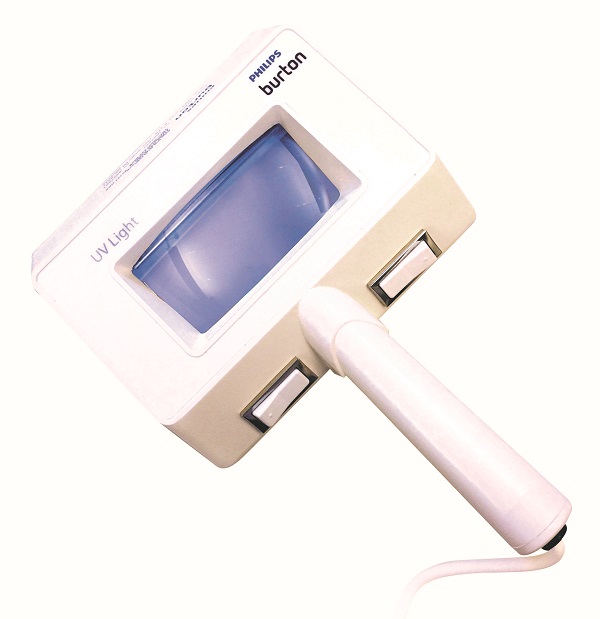
What is a Woods Lamp?
A Woods lamp is a low-output mercury arc that emits a wavelength 320-450 nm. True to its name, it was invented by Baltimore physicist Robert W. Wood in 1903 and was first used within the discipline of dermatology in order to detect fungal infections of hair. Woods lamps are sometimes incorrectly referred to as wood’s lamps.
The ultraviolet light the Woods lamp emits is undetectable by the naked eye; however, it glows violet within a dark setting because it also emits light from the violet section of the electromagnetic spectrum.
A traditional Woods lamp package may include a variety of phosphors with a selection of peak emissions, a magnifying lens, both a white light and a dark light, and a heavy cloth to help exclude all visible light. Woods lamps are available in a variety of models to help meet the needs of your health care facility.
A portable Woods lamp is small, durable, easy to use, safe, and inexpensive–making it a sound investment for your medical practice. In addition, a Woods lamp skin analysis can provide rapid results, which can be a tremendous help in providing a timely and reliable patient diagnosis.
What is Fluorescence?Fluorescence describes the colored glow that is visible when some substances – including porphyrins and collagen – absorb black light and then re-emit it at a longer wavelength within the visible spectrum. The properties of fluorescence allow many skin conditions to visibly stand for a quick visible diagnosis by your health care team. Woods lamp examination can either support or refute a suspected diagnosis, based on the colors of fluorescence displayed by the affected skin being examined. In addition, a Woods lamp will allow your team to see subtle changes in color, even if they are invisible to the naked eye.
Top 9 Medical Diagnostic Applications for a Woods Lamp
Normal, healthy skin looks purple or violet during a Woods lamp examination and will not glow. This allows fungal and bacterial colonies to stand out since they will naturally fluoresce under the ultraviolet light.
Depending on the skin condition revealed, fluorescence can appear as golden yellow, pale green, aqua, pink-orange, blue-white, or even purple-brown.
1. Tinea Capitis
Tinea capitis describes a condition in which the skin presents with areas of scale and baldness as a result of a fungal infection. It was the first skin condition for which the Woods lamp was historically used to diagnose. Woods lamp diagnosis is not only effective for identifying individual cases of tinea capitis, but also for detecting and controlling large epidemics, such as within schools. However, it is important for your team to keep in mind that tinea capitis fluoresces in only 5 percent or fewer of cases in the United States. When it does, it presents typically as blue-green, although it can occasionally manifest as dull yellow or dull blue. However, other fungal infections related to different microorganisms do not necessarily fluoresce.
2. Vitiligo
A Woods lamp examination is especially helpful in making a diagnosis of vitiligo – especially if it must be differentiated from other conditions like leprosy, pityriasis or post-inflammatory hypopigmentation. The lamp also helps illuminate evolving lesions in a fair-skinned patient. Vitiligo appears as white patches under a Woods lamp, which varies in both location and extent. These patches may also spread and grow over time. Examining under a Woods lamp can give your health care team complete visual information regarding the progress of a patient’s vitiligo.
3. Corneal Abrasions
To look for scratches on the eye, the medical professional must first put a fluorescein solution in the patient’s eye before using the Woods lamp to illuminate the eye area – any scratches or abrasions will subsequently glow when the ultraviolet light hits them. During an eye exam, a Woods lamp can help identify foreign particles within the eye, even glass and other hard-to-see particles, plus illuminating injuries and even blocked tear ducts.
4. Ringworm
This fungal culture, which often affects the skin, hair, and nails, is easily detected and/or confirmed using a Woods lamp. The use of a Woods lamp to test for ringworm is especially common within veterinary practices, as the infection presents often among animals. Under the Woods lamp, the ringworm infection fluoresces as bright apple green.
5. Porphyria
Porphyria is a group of inherited disorders that cause rashes, blistering, and scarring of the skin. When examined under a Woods lamp, they all fluoresce as red-pink. In fact, detecting and identifying excess porphyrins in urine, teeth, red blood cells, stool samples and even blister fluid can easily be achieved when using a Wood’s lamp.
6. Pigment Disorders
Hypopigmentation or depigmentation can be difficult to discern in the fairest-skinned of patients, but they demonstrate increased border sharpness when examined using a Woods lamp; they generally fluoresce as a bright blue-white because of the increased amount of dermal collagen being illuminated, which is a result of either decreased or absent intervening melanin. Conversely, hyperpigmentation presents with enhanced border contrast under a Woods lamp because of the increased absorption of light related to increased levels of melanin. Using a Woods lamp can help determine the level of melanin within the skin, along with clearly highlighting variations in epidermal pigmentation.
7. Bacterial Infections
Infections from pathogens like Pseudomonas can appear as a bright green fluorescence under a Woods lamp. This method is often used to examine burn wounds for bacteria and also is helpful in the diagnosis of Stevens-Johnson syndrome.
8. Acne Vulgaris
Orange-red fluorescence is associated with any comedones infected with P. acnes. This can help the health care professional determine which, if any, comedones are the result of infection.
9. Other Miscellaneous Uses
- Other uses of the Woods lamp in health care include the following:
- Detection of systemic drugs like tetracycline
- Efficacy assessment of the sun protection factor of various sunscreens on the market
- Detection of skin allergens
- Calculation of circulation time
- Treatment of warts, with some success
- Detection and treatment of skin cancer, with some success
How to Use a Woods Lamp
A Woods lamp is relatively simple to use, but it does require a few important steps to make sure results are valid. For example, your dermatologist should advise all patients undergoing any type of Woods lamp test to avoid washing the area to be tested before undergoing the diagnostic test. They should also avoid using any medications, deodorant, perfume, and makeup on the testing area – some product ingredients can cause the skin to change color under the light, which can influence test results, providing in either a false negative or a false positive.
In addition, a Woods lamp works best in a completely dark environment – your team should turn off all lights and draw any curtains or window shades in the room. Some physicians even use black drapes or light-canceling shades to ensure utter darkness. The Woods lamp typically takes about one minute to warm up, and then it’s ready for use.
The user should aim the light directly above the sample or lesions being studied, carefully inspecting the area and paying attention to all zones that fluoresce and noting their borders – ideally the medical professional will hold the light around 4-5 inches from the skin to look for color changes.
Your team should watch out for any topical medications, soap residue, lint, etc., which can create a false positive that typically presents as red-pink. If they notice any of these complicating factors, they can easily be removed. Healthy skin will show as slightly blue, but the physician may also see white spots where skin is thickened, yellow, oilier spots and purple spots where skin is dehydrated.
Woods Lamp Safety
The black light a Woods lamp emits is harmless to the patient, as it does not emit short-wavelength ultraviolet radiation. A Woods lamp cannot cause sunburn or sun damage–or in any other way degrade healthy skin, except in rare cases of patients with extreme photosensitivity. In these cases, the patients run the risk of developing a rash on areas of the skin exposed to the UV light. However, out of an abundance of caution, it is considered a best practice to ask the patient to close his/her eyes when using the Woods lamp to examine the face – one should also avoid shining the light directly in the patient’s eyes.